baby chest x ray technique
Chest x-ray technique reduces radiation dose in infants. When the chest radiograph also includes the abdomen look out for the umbilical clip.

Chest X Ray For Students How To Interpret And Present Methodically Andreas Astier
The chest radiograph is the most common radiographic procedure.

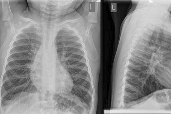
. The normal neonatal chest X-ray Aerationofthenormalneonatallungisvirtuallycomplete within two or three respiratory cycles. Make use of digital radiography dr and needle phosphor computerised. First of all have a look to see if the neonate is premature or not - signs of prematurity being reduction in subcutaneous fat and the lack of humeral head ossification.
Inspiration Penetration Rotation is part of the Lecturio course Radiology WATCH the complete course on httplect. Baby chest x ray technique. X-ray plate - avoid direct contact with baby use X-ray tray provided with the open care system cover with sterile plastic sheetenvelope Avoid direct contact with the cold X-ray plate Provide.
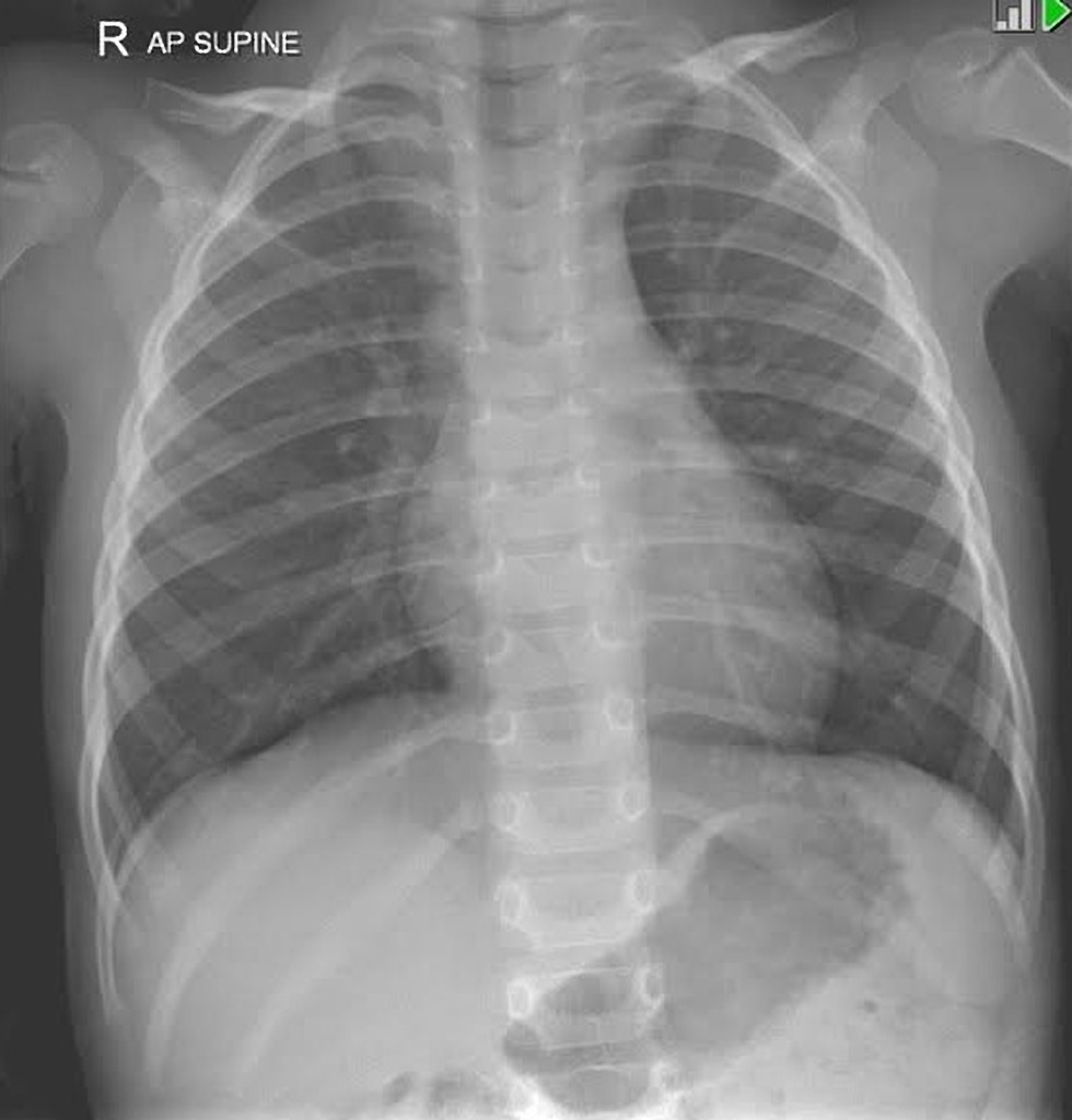
Erect chest X-rays are taken at 180 cm. In this study the exposure technique of 65 kVp and 16 mAs was chosen as a reference image due to this technique being near the suitable exposure uses in pediatric chest. These are plastic clips used to clamp the umbilicus before it is cut at birth.
Full legfull spine imaging is performed at 180 cm using CR. August 26 2021 -- X-ray dark-field chest. This video Chest X-Ray Techniques.
Baby chest x ray technique. Pediatric Chest Screen 70-80 DIGITAL OPTIMUM kVp Universal CR Technique Chart using a standard 21 LgM Part View kV mAs kV mAs kV mAs Abdomen AP Grid 85 10 -15 85 20 - 25. Lateral cervical spines are taken at 150 cm.
Insufficient inspiration To overcome these a variety of techniques can be used.

Pedia Poser For Xray Imaging

The Normal Cxr Radiology Nurse Respiratory Therapy
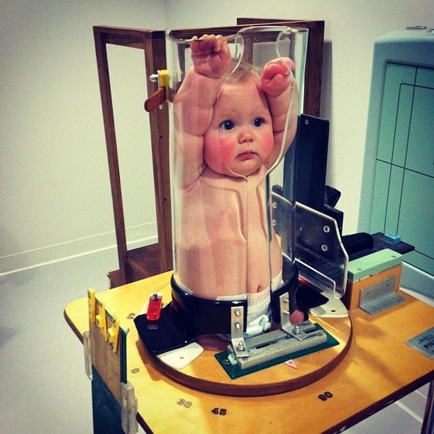
Ce4rt Guide For X Ray Techs To Immobilize Pediatrict Patients

Pin On Raising Healthy Congenital Diaphragmatic Hernia Children

Pin On Xrays

Diagnosis Of Other Lung Conditions In Premature Babies

Chest X Ray Multiple Bilateral Rib Fractures In Various Stages Of Download Scientific Diagram
![]()
Normal Chest X Ray Anatomy Tutorial Kenhub
Normal Chest Radiograph Pediatric Radiology Case Radiopaedia Org

Pin By Heather Royal On School Daze Diagnostic Imaging Radiology Schools Med Student

Xrays Pdf Dramatic Play Preschool Kids Vet Clinic Dramatic Play Centers

T Spine Image Radiology Imaging Radiology Student Radiology

Pediatric Chest Supine View Radiology Reference Article Radiopaedia Org
Chest X Ray Portable Upright Semi Upright Supine Positioning And Technique Youtube
Choosing Wisely At Sickkids Means Holding Off On X Raying Infant Bronchiolitis Cases Coming To The Ed
2

Neonatal Chest Radiograph In The Exam Setting Radiology Reference Article Radiopaedia Org

Chest X Ray Appearances In Ventilated Infants With Mas A Typical Download Scientific Diagram

Magnets May Pull Kids With Sunken Chests Out Of Operating Room Pectus Excavatum Chest Marfan Syndrome